Our Health Library information does not replace the advice of a doctor. Please be advised that this information is made available to assist our patients to learn more about their health. Our providers may not see and/or treat all topics found herein.
Extremity X-Ray
Test Overview
An extremity X-ray is a picture of your hand, wrist, arm, foot, ankle, knee, hip, or leg. It is done to see whether a bone has been fractured or a joint dislocated. It is also used to check for an injury or damage from conditions such as an infection, arthritis, bone growths (tumors), or other bone diseases, such as osteoporosis.
Why It Is Done
Extremity X-rays are done to:
- Find the cause of pain in an extremity.
- See if your bone is fractured or your joint is dislocated.
- See if fluid has built up in the joint or around a bone.
- See if your bones are positioned properly after treatment for a fracture or dislocation, such as after placing a cast or splint on an arm or leg. An X-ray also may be done after a doctor places a device such as a pin or an artificial joint in a bone.
- Find changes in your bones caused by conditions such as an infection, arthritis, bone growths (tumors), osteoarthritis of the hip, osteoarthritis of the knee, or other bone diseases.
- Find foreign objects such as pieces of glass or metal.
- Check to see if a child's bones are growing normally.
- See if your bones and joints are in the correct position after joint replacement surgery.
How To Prepare
In general, there's nothing you have to do before this test, unless your doctor tells you to.
How It Is Done
You will need to remove any jewelry that may be in the way of the X-ray picture. You may need to take off some of your clothes, depending on which area is examined. You will be given a cloth or paper gown to use during the test. You may be allowed to keep on your underwear if it does not get in the way of the test.
During the test
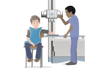
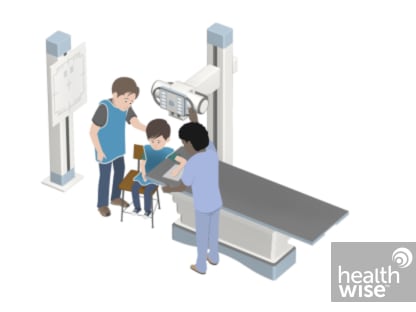
During the X-ray test, you will sit by or be on an X-ray table. The X-ray technologist will position your limb. If you have an injury, your leg or arm will be handled gently and supported when moved or repositioned. Pillows, sandbags, or other objects may be used to hold the injured limb in place while the pictures are taken. If you are wearing a brace or other device, it may need to be removed. A lead shield may be placed over your pelvic area to protect it from radiation.
Two or more pictures of the affected limb are usually taken. X-ray pictures may also be taken of other joints or limbs, since an injury at one point may cause damage somewhere else. Sometimes an X-ray picture of the unaffected limb is taken so it can be compared with the affected limb.
How long the test takes
An extremity X-ray usually takes about 5 to 10 minutes. You will wait about 5 minutes until the X-rays are processed, in case repeat pictures need to be taken. In some clinics and hospitals, X-ray pictures can be shown right away on a computer screen (digitally).
Watch
How It Feels
You won't feel any pain from the X-ray itself. You may find that the positions you need to hold are uncomfortable or painful. This is more likely if you have an injury.
Risks
There is always a slight chance of damage to cells or tissue from radiation, including the low levels of radiation used for this test. But the chance of damage from the X-rays is extremely low. It is not a reason to avoid the test.
If you need an X-ray during pregnancy, a lead apron will be placed over your belly to protect the baby from exposure to radiation from the X-rays. The chance of harm is usually very low compared with the benefits of the test.
Results
In an emergency, the doctor can see the initial results of an extremity X-ray in a few minutes. Otherwise, a radiologist usually has the official X-ray report ready the next day.
Normal results:
- The bones, joints, and soft tissue look normal. No foreign objects, such as fragments of metal or glass, are present.
- No infection and no abnormal growths (tumors) are present.
- The joints are normal with no dislocation or signs of disease, such as arthritis.
- All parts of a joint replacement are in the correct position.
Abnormal results:
- Fractured bones may be present.
- Foreign objects, such as fragments of metal or glass, may be present.
- Abnormal growths (tumors) are present.
- Signs of bleeding or infection, such as a collection of blood, pus, or gas may be present.
- A joint may be dislocated.
- The bones or joints may show signs of damage from a disease such as osteoporosis, osteoarthritis, gout, Paget's disease, or rheumatoid arthritis of the feet and hands.
- Swelling is present in tissues around the bones even though the bones may be normal.
- There are loose parts, worn parts, or an infection in a joint that has artificial pieces (joint replacement).
Related Information
Credits
Current as of: March 26, 2025
Author: Ignite Healthwise, LLC Staff
Clinical Review Board
All Ignite Healthwise, LLC education is reviewed by a team that includes physicians, nurses, advanced practitioners, registered dieticians, and other healthcare professionals.
Current as of: March 26, 2025
Author: Ignite Healthwise, LLC Staff
Clinical Review Board
All Ignite Healthwise, LLC education is reviewed by a team that includes physicians, nurses, advanced practitioners, registered dieticians, and other healthcare professionals.
This information does not replace the advice of a doctor. Ignite Healthwise, LLC disclaims any warranty or liability for your use of this information. Your use of this information means that you agree to the Terms of Use and Privacy Policy. Learn how we develop our content.
To learn more about Ignite Healthwise, LLC, visit webmdignite.com.
© 2024-2026 Ignite Healthwise, LLC.


